Friday, September 06, 2019

Mesh electronics could make brain stimulation the new therapeutic norm

Implantable brain electrodes have been around for quite some time now, both for diagnosing and treating neuropsychiatric conditions like Parkinson’s disease. However, one limitation of conventional probes is their size and rigidity, compared to the soft, gelatinous consistency of the brain.

A new research paper offers a revolutionary solution: mesh electronics, which allows personalized electronics that mimic the neuron’s shape, size and texture, to be created for any brain-related disorder.
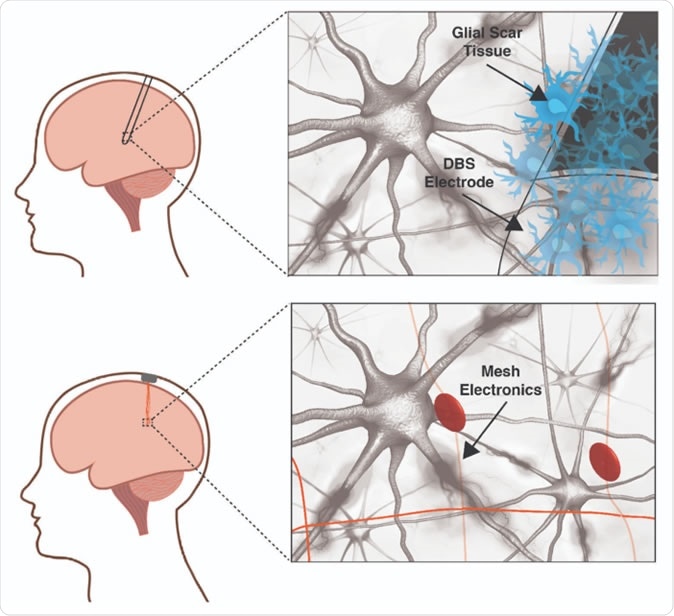
A traditional deep brain stimulation electrode (top panel) provokes an immune response in the brain while a mesh electronic interface (bottom panel) does not. The size and rigidity of the DBS electrode result in chronic inflammation causing glial scarring between brain tissue and electrode, degrading the neural interface. Mesh electronics evade the immune response due to cellular and sub-cellular features and bending stiffness resembling the brain itself. Image courtesy of Shaun Patel and Charles Lieber
A traditional deep brain stimulation electrode (top panel) provokes an immune response in the brain while a mesh electronic interface (bottom panel) does not. The size and rigidity of the DBS electrode result in chronic inflammation causing glial scarring between brain tissue and electrode, degrading the neural interface. Mesh electronics evade the immune response due to cellular and sub-cellular features and bending stiffness resembling the brain itself.

Why are implantable electrodes needed?

All neuroscientists agree that the foundation of cognition is the brain, including thinking, emotions, memory, judgment and decision-making. The same applies to all kinds of brain disorders, according to researcher. She became interested in this field after witnessing a 500 ms pulse of electricity conferring an impulsive risk-taker with the momentary ability to make a safer choice, without even knowing how it occurred. The alteration of cognitive thinking, in other words, happened below the level of consciousness.

However, it is difficult for scientists to trace behaviors to specific neurons or brain areas that are dysfunctional, though this could help discover where illnesses such as addiction, Alzheimer’s or Parkinson’s disease originate. Therapy for these conditions is currently limited to drugs and electrical stimulation via implantable electrodes.

Medical treatment is mostly with neuroactive drugs like L-dopa in Parkinson’s disease, which stills the quivering movements that make voluntary actions so difficult. However, the off-target or nonselective inhibition of L-dopa actions causes severe and even unacceptable side effects, on various body systems, including nausea, depression or arrhythmia.

The failure of drug treatment is an indication for electrode implantation to provide Deep Brain Stimulation (DBS). These work by sending automated pulses of electrical energy to paired implants, each about the size of a pencil, which is huge with respect to the brain. These FDA-approved electrodes must be implanted while the patient is awake and able to guide the surgeons during their calibration of the required current intensity that will stop the tremors.

Describing the effect of DBS, researcher  marvels, “Almost instantly, you can see the person regain control of their limbs.” However, these electrodes aren’t quite foolproof either, causing inadvertent stimulation of other brain areas at times, which causes disturbing adverse effects such as speech impediments. Just as bad, the brain has an active and vigilant immune system composed of neuroglial cells, which eventually disrupts the electrode’s efficient functioning by smothering it in a protective glial covering. This may also grow so large as to mechanically affect or even kill neighboring neurons. The limited device life caused by unnecessary continuous operation, and the lack of feedback which hampers operational efficiency, are other limitations. While many of these are being individually overcome, the rigid and imprecise neural interface is a basic restriction on further progress.
Implantable rigid electrodes vs mesh electronics

The increased rigidity of even the softest conventional probes causes several problems: chronic inflammation and scarring, mismatched stiffness which leads to a movement of the probe and neuron away from each other relatively quickly, and hinders tracking of the same neuron or circuit; and the solid probe lies uneasily on the quite differently shaped brain surface. This topological difference pushes neurons away from their rightful place, while also preventing repair of broken synaptic connections and blocking the free movement of chemicals in the brain environment.
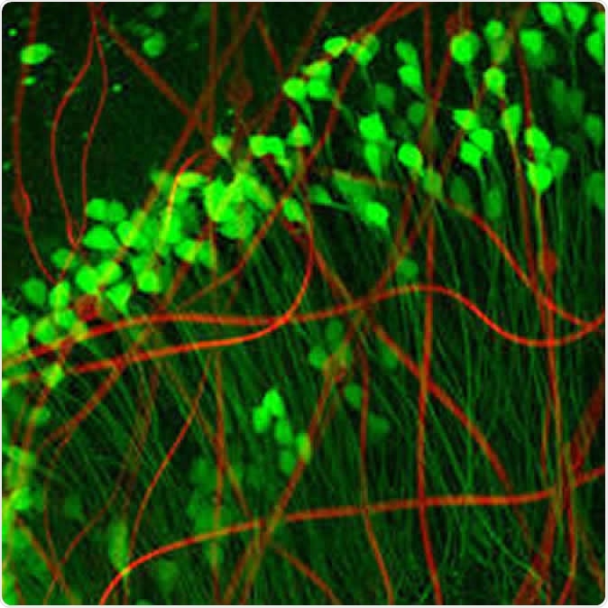
Neuron-like electronics (red) mimic the shape, size, and flexibility of neurons (green), enabling them to maintain symbiosis with native brain tissue (Credit: Xiao Yang, Lieber Lab)
Neuron-like electronics (red) mimic the shape, size, and flexibility of neurons (green), enabling them to maintain symbiosis with native brain tissue
This is where Lieber has made a huge difference with ultra-flexible mesh electronics, which promise to deliver what he calls “precision electronic medicine”. These hardly activate an immune response, but remain very close to the cells they are intended to spy on. As such, they offer a way to obtain reliable information on the communication between individual neurons or its breakdown. This could help elucidate cross-talk between specific subsets of neurons. It is also necessary if any degree of precise regulation is to be achieved via feedback circuits, which requires a two-way transmission of information through the neural device. All this could produce a more accurate and detailed map of the brain circuitry. This could potentially open the door to the successful treatment of any brain disorder.
Moreover, Lieber’s mesh electrodes have a peculiar benefit in their ability to guide developing neurons to the right place, such as injured areas as following a stroke. They could potentially encourage directed neuronal migration to rebuild injured or degenerated areas, and integrate these cells into the right circuits, using differentially modulated successive pulses of electricity.

The risk – and the future

Lieber envisages other mind-blowing advances: special electrodes could be designed to provide precise control over prosthetic or paralyzed limbs, and this could serve as an efficient substitute for damaged brain connections, as well as autocalibration of the signal based on live feedback. He says, “If you could actually interact in a precise and long-term way and also provide feedback information, you could really communicate with the brain in the same way that the brain is communicating within itself.”

Other major would-be players in this field include Neuralink from Elon Musk, which aims at assisting paralyzed patients by providing a mind-computer interface so they can use their computers; Facebook which is looking at texting via word imaging; and Kernel from Brian Johnson, which is focused on increasing cognitive abilities. Lieber emphasizes that his desire is to help people who are sick – and even to prevent the aging-related decline in memory if possible. However, strict ethical control is mandatory lest such mind-altering power be misused. While the benefits are obvious, such as helping people overcome addiction or obsessive-compulsive disorder, the downside is equally plain – the patient’s thoughts are laid open to scrutiny, despite being the innermost stronghold of a person. Secondly, it raises the risk of putting memory, learning ability and other  desirable mental traits up on sale, for people who would like to enhance their brain capability.

Meanwhile, scientists like Lieber and Patel continue to refine mesh electronics, by increasing the number of electrodes that can be implanted, designing live feedback systems to enable continuous recalibration, and improving the processing of the voluminous data collected by the electrodes. They urge a blending of multiple disciplines to help advance neurotechnology to the point where it breaches the limit of machine-mind interface. Patel says, “The next frontier is really the merging of human cognition with machines.”

this is only for your information, kindly take the advice of your doctor for medicines, exercises and so on.   
https://gscrochetdesigns.blogspot.com. one can see my crochet creations 
https://gseasyrecipes.blogspot.com. feel free to view for easy, simple and healthy recipes    
https://kneereplacement-stickclub.blogspot.com. for info on knee replacement


Labels: , , , , , , ,

Saturday, March 23, 2019

Doctors Wired a Prosthetic Hand Directly Into a Woman’s Nerves

In a world first, doctors in Sweden  a prosthetic hand directly into a woman’s nervesallowing her to move its fingers with her mind and even feel tactile sensations.

The hand is an enormous step up from existing prostheses, which often rely on electrodes placed on the outside of the skin — and it could herald a future in which robotic devices interface seamlessly with our bodies.

Nerve Case

Researchers created the prosthetic hand as part of DeTOP, an ambitious European research program on prosthetic limbs.

Surgeons anchored the hand to the woman’s forearm bones using titanium implants. They connected an array of 16 electrodes directly to her nerves and muscles, allowing her to control the hand with her mind — and, according to photos, use it to tie shoelaces and type on a laptop computer.
“The breakthrough of our technology consists on enabling patients to use implanted neuromuscular interfaces to control their prosthesis while perceiving sensations where it matters for them, in their daily life,” said the researchers.

Electronics wired straight into a human nervous system allow for mind-bending new ways to interact with technology. A video released by the Swedish researchers even shows the woman using the implant to flex a virtual hand on a computer screen — before the actual physical hand was installed.
For decades, cyborg limbs like those depicted in “Star Wars” or “Neuromancer” seemed relegated to the realm of science fiction. New research shows that they’re already here — just not yet widely available.

THIS IS ONLY FOR INFORMATION, ALWAYS CONSULT YOU PHYSICIAN BEFORE HAVING ANY PARTICULAR FOOD/ MEDICATION/EXERCISE/OTHER REMEDIES.                                    PS- THOSE INTERESTED IN RECIPES ARE FREE TO  VIEW MY BLOG-                                                                                           https://gseasyrecipes.blogspot.com/                                                                                                                                                         FOR INFO ABOUT KNEE REPLACEMENT, YOU CAN VIEW MY BLOG-                                                  https:// kneereplacement-stickclub.blogspot.com/           

                   FOR CROCHET DESIGNS                                                                                                                                                                                                  https://gscrochetdesigns.blogspot.com


Labels: , , , , , , , , , , , , , , ,

Tuesday, February 05, 2019

Novel technique could provide early lung cancer diagnosis

Researchers have developed a new technique that has the potential to identify specific lung cancer markers at the earliest possible stage in a convenient and reusable way.

The new technique could create a highly sensitive graphene biosensor with the capability to detect molecules of the most-common lung cancer bio-markers.

In addition, the new biosensor design could revolutionise the existing electronic nose (e-nose) devices that identify specific components of a specific vapour mixture — a person’s breath — and analyses its chemical make-up to identify the cause.

“The new biosensors which we have developed show that graphene has significant potential for use as an electrode in e-nose devices.

“For the first time, we have shown that with suitable patterning graphene can be used as a specific, selective and sensitive detector for bio-markers,” said a researcher.

With further development of the devices, a cheap, reusable and accurate breath test for early-stage detection of lung cancer can become a reality, the researchers added in a paper published recently.

Lung cancer is one of the most common and aggressive cancers, killing around 1.4 million people worldwide each year.

THIS IS ONLY FOR INFORMATION, ALWAYS CONSULT YOU PHYSICIAN BEFORE HAVING ANY PARTICULAR FOOD/ MEDICATION/EXERCISE/OTHER REMEDIES.                                    PS- THOSE INTERESTED IN RECIPES ARE FREE TO  VIEW MY BLOG-                                                                                           https://gseasyrecipes.blogspot.com/       
                                                                                                                                                                      FOR INFO ABOUT KNEE REPLACEMENT, YOU CAN VIEW MY BLOG-                                                  https:// kneereplacement-stickclub.blogspot.com/           
                                                                         
  FOR CROCHET DESIGNS                                                                                                                                                                                                  https://gscrochetdesigns.blogspot.com

Labels: , , , , , , , , , , , ,

Thursday, June 14, 2018

This Neural Jelly-Like Implant is the Future

While brain-analyzing technologies such as MRI scans are ever-improving, sometimes researchers need to get inside their subjects’ brain in a more literal sense. For example, in order to get readings from specific brain cells, as well as to integrate brains with prosthetic or medical devices, a physical electrode must be inserted into the brain itself.
 
As one can imagine, this isn’t fun at all. In fact, recently, scientists have called for improved safety guidelines and better research into how these electrodes destroy brain cells, as well as how they trigger the brains’ inflammatory immune system response.
 
Now, researchers  may have found a way around this. They’ve developed a flexible, squishy-silicon-based hydrogel that sticks to neural tissue, bringing non-invasive electrodes to the brain’s surface.

This hydrogel, which was tested on a cat’s dorsal root ganglion – a cluster of spinal nerve cells – can conform and adhere to cells. This lets the enclosed electrodes record brain activity without actually exposing the brain to the electrodes.

The researchers are hoping that their work will lead to a new era of safer neural implants, while also giving rise to better, more accurate neural readings that could help us better understand diseases and other brain conditions.

In a press release, an engineer who worked on the hydrogel said: “Imagine you have a bowl of Jell-O, and you insert a rigid plastic fork into the bowl and move it around.” This is what it’s like when a conventional electrode is inserted into the brain. 

While this is an over exaggeration, electrodes do cause a tiny amount of structural damage on their way down through the cortex. While scientists are still split on whether or not the adult human brain can grow new cells, it’s probably best to minimize the number of holes that you drill into it.

Furthermore, the brain knows that electrodes don’t belong there, so it calls on the immune system to fight them off. This leads to the eventual degradation of electrodes, meaning they give off inaccurate cell recordings and hamper neuroscientific research. Since the hydrogel can pass as brain-like, the body is much less likely to try to fight it off.

It’s far too early to say exactly how this new neural implant hydrogel will affect the realm of medical neuroscience, but if human trials work as well as the experiments on cats, then the brain-computer interfaces of the future could become much more seamless and a lot less harmful.
THIS IS ONLY FOR INFORMATION, ALWAYS CONSULT YOU PHYSICIAN BEFORE HAVING ANY PARTICULAR FOOD/ MEDICATION/EXERCISE/OTHER REMEDIES.                                                                                                                                                                                                        PS- THOSE INTERESTED IN RECIPES ARE FREE TO  VIEW MY BLOG-                                                                                           https://gseasyrecipes.blogspot.com/  

FOR INFO ABOUT KNEE REPLACEMENT, YOU CAN VIEW MY BLOG-                                                                                        https:// kneereplacement-stickclub.blogspot.com/                              
FOR CROCHET DESIGNS                                                                                                                                                                                                                                 https://my crochet creations.blogspot.com

Labels: , , , , , , , , , , , , , , ,

Thursday, May 03, 2018

Brain implant gives patients control over Parkinson's disease

Parkinson's disease can be challenging even in its early stages.

But one Texas woman has been able to regain control of her life.

She was diagnosed with the neurological disease five years ago at age 40.

Her tremors and muscle stiffness got so bad, she couldn't cook or even wash her hair.

So, last October she decided on deep brain stimulation.

"A lot of times, these patients are trapped in their bodies because they can't do things they want to do," said the Dr. "This surgery allows them to go back to a more normal life."

During the surgery, electrodes are implanted into the brain while another device, like a pacemaker, goes near the collarbone. It sends impulses to the brain which block the abnormal nerve signals that cause tremors.

THIS IS ONLY FOR INFORMATION, ALWAYS CONSULT YOU PHYSICIAN BEFORE HAVING ANY PARTICULAR FOOD/ MEDICATION/EXERCISE/OTHER REMEDIES.                                                                                                                                                                                                        PS- THOSE INTERESTED IN RECIPES ARE FREE TO  VIEW MY BLOG-                                                                                           https://gseasyrecipes.blogspot.com/  

FOR INFO ABOUT KNEE REPLACEMENT, YOU CAN VIEW MY BLOG-                                                                                                                              https:// kneereplacement-stickclub.blogspot.com/                              
FOR CROCHET DESIGNS                                                                                                                                                                                                                                 https://my crochet creations.blogspot.com
  

Labels: , , , , , , , , , , ,

Tuesday, December 20, 2016

An Electronic Implant to Return Motor Function

There’s renewed hope that paraplegic and quadriplegic patients will one day be able to return motor and sensory function to their affected limbs, thanks to a brand-new invention by doctors in the US.
 
The stimulator they have come up with bypasses spinal injuries by forcing the body to use alternative neural pathways in order to transmit signals to the body’s extremities. In fact, the most recent test that the doctors conducted saw a quadriplegic’s finger motion improve by 300% while simultaneously improving grip strength, giving the patient the ability to perform everyday tasks once again.

Last June, the doctors implanted the stimulator into the body of a 28-year-old quadriplegic named Brian Gomez. The operation was conducted at Ronald Reagan University in California, USA. Gomez suffered his injuries during a dirt biking accident when he was 23 years old.

He broke his neck at his C-5 vertebra, which is at the middle of the neck next to the thyroid cartilage. This means that he lost all motor functions at his extremities, but retained head-to-toe sensation. Due to the nature of the injury he suffered, he was considered to be the perfect candidate for the experimental treatment.
Gomez assisted the researchers by giving them feedback on the sensations he was feeling from the stimulator so that they could be adjusted accordingly. The stimulator contains a series of 32 electrodes that can send pulses around the injured area, and that’s why it was implanted into the patient’s neck – to bypass the injury.
A great analogy for how the stimulator works is what occurs when there’s an accident on a freeway. While it’s almost inevitable that traffic will come to a standstill, there are numerous side streets and detours that can be taken to bypass the scene of the accident and reach the final destination.
 Neural signals are like traveling vehicles, the scene of the accident is the injury, and the alternate routes are the new pathways that the stimulator helps the spinal cord to find.
 
In addition to the stimulator and its electrodes, the researchers also implanted a battery pack into the patient so that the electrical stimulation they were providing to him could be fine-tuned into certain patterns, thus providing more or less stimulation to certain areas as needed.
The concept behind this is to stimulate neural pathways that the brain might be ignoring. Over time, these pathways open up and allow messages to pass through them. In essence, the whole process is like retraining the body to use the undamaged “side streets” so that the neural messages can arrive, thus returning functionality to the body’s extremities.

This particular study marks the very first time that doctors have tried electrode technology on someone suffering from quadriplegia stemming from a neck injury. It’s also the first time that such technology has been implanted under the skin to attempt to treat such an injury.

Despite the excitement resulting from this genuine medical breakthrough, it must be emphasized that the researchers’ intention was never to return affected limbs back to 100% functionality. In fact, the main goal was for hands to be able to impart enough force in order for the patient to be able to do tasks such as brushing his teeth, tying his shoelaces, or simply eating food with a fork.

Although the breakthrough is undoubtedly fascinating, researchers are exercising caution with regard to expectations, and this is because the stimulator has only been tested on a single patient to date. More data needs to be gathered, followed by a peer review and publication of the researchers’ definitive results. 

Nevertheless, it’s a hugely exciting time for paraplegics and quadriplegics who now have hope of returning some motor function to their extremities.

this is only for your information, kindly take the advice of your doctor for medicines, exercises and so on.
https://gscrochetdesigns.blogspot.com. one can see my crochet creations
https://gseasyrecipes.blogspot.com. feel free to view for easy, simple and healthy recipes 
https://kneereplacement-stickclub.blogspot.com. for info on knee replacement

Labels: , , , , , , , , , , , ,

Wednesday, September 30, 2015

New brain implant to treat people with memory loss

THIS IS ONLY FOR INFORMATION, ALWAYS CONSULT YOU PHYSICIAN BEFORE HAVING ANY PARTICULAR FOOD/ MEDICATION/EXERCISE/OTHER REMEDIES.


PS- THOSE INTERESTED IN RECIPES ARE FREE TO VIEW MY BLOG-
HTTP:GSEASYRECIPES.BLOGSPOT.COM


FOR INFO ABOUT KNEE REPLACEMENT, YOU CAN VIEW MY BLOG
HTTP://KNEE REPLACEMENT-STICK CLUB.BLOGSPOT.COM/

FOR CROCHET DESIGNS- http://My Crochet Creations.blogspot.com/
 
 Researchers have developed a brain implant that translates short term memories into longer-term ones and may help dementia sufferers and brain-damaged patients retain their memory. 
 
Researchers at the University of Southern California and Wake Forest Baptist Medical Center said the prosthesis, which includes a small array of electrodes implanted into the brain, has performed well in laboratory testing in animals and is currently being evaluated in human patients.

Designed originally at USC and tested at Wake Forest Baptist, the device builds on decades of research by Ted Berger and relies on a new algorithm created by Dong Song, both of the USC Viterbi School of Engineering.

The development also builds on more than a decade of collaboration with Sam Deadwyler and Robert Hampson of the Department of Physiology & Pharmacology of Wake Forest Baptist who have collected the neural data used to construct the models and algorithms.

When brain receives the sensory input, it creates a memory in the form of a complex electrical signal that travels through multiple regions of the hippocampus, the memory centre of the brain.
At each region, the signal is re-encoded until it reaches the final region as a wholly different signal that is sent off for long-term storage. If there's damage at any region that prevents this translation, then there is the possibility that long-term memory will not be formed.

That's why an individual with hippocampal damage (for example, due to Alzheimer's disease) can recall events from a long time ago - things that were already translated into long-term memories before the brain damage occurred - but have difficulty forming new long-term memories.

Song and Berger found a way to accurately mimic how a memory is translated from short-term memory into long-term memory, using data obtained by Deadwyler and Hampson, first from animals, and then from humans.

Their prosthesis is designed to bypass a damaged hippocampal section and provide the next region with the correctly translated memory.

In experiments, Hampson and Deadwyler read the electrical signals created during memory formation at two regions of the hippocampus in patients who had electrodes implanted in their hippocampi to treat chronic seizures.
 
The information was then sent to Song and Berger to construct the model.

The team then fed those signals into the model and read how the signals generated from the first region of the hippocampus were translated into signals generated by the second region of the hippocampus.

In hundreds of trials conducted with nine patients, the algorithm accurately predicted how the signals would be translated with about 90 per cent accuracy.

"Being able to predict neural signals with the USC model suggests that it can be used to design a device to support or replace the function of a damaged part of the brain," Hampson said.

Labels: , , , , , , ,