Everything one must know about bladder cancer
What Is Bladder Cancer?
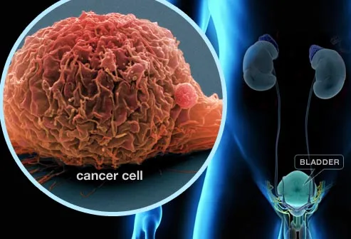
Cancer is the growth of abnormal cells in the body. Bladder cancer typically begins in the inner lining of the bladder, the organ that stores urine after it passes from the kidneys. Most bladder cancers are caught early, when treatments are highly successful and the disease has not spread beyond the bladder. But bladder cancer tends to come back, so regular check-ups are important.
Warning Sign: Blood in Urine
Blood in the urine can be a sign of bladder cancer, either visible to the eye or picked up by routine testing. The urine may look darker than usual, brownish, or (rarely) bright red. Most commonly, blood in the urine is not caused by cancer, but by other causes. These include exercise, trauma, infections, blood or kidney disorders, or drugs, such as blood thinners.
Cancer is the growth of abnormal cells in the body. Bladder cancer typically begins in the inner lining of the bladder, the organ that stores urine after it passes from the kidneys. Most bladder cancers are caught early, when treatments are highly successful and the disease has not spread beyond the bladder. But bladder cancer tends to come back, so regular check-ups are important.
Warning Sign: Blood in Urine
Blood in the urine can be a sign of bladder cancer, either visible to the eye or picked up by routine testing. The urine may look darker than usual, brownish, or (rarely) bright red. Most commonly, blood in the urine is not caused by cancer, but by other causes. These include exercise, trauma, infections, blood or kidney disorders, or drugs, such as blood thinners.
Warning Sign: Bladder Changes
Bladder symptoms are more likely to come from conditions other than cancer. But bladder cancer can sometimes cause changes to bladder habits, including:
Needing to go, with little or no results
Having to go more often than usual
Painful urination
Difficulty urinating
Urinary tract infections or bladder stones can cause similar symptoms, but require different treatments.
Smoking
Although the exact causes of bladder cancer remain unknown, smoking is the leading risk factor. Smokers are about four times more likely to get bladder cancer than people who have never smoked. Chemicals in tobacco smoke are carried from the lungs to the bloodstream, then filtered by the kidneys into urine. This concentrates harmful chemicals in the bladder, where they damage cells that can give rise to cancer.
Chemical Exposure
Research suggests that certain jobs may increase your risk for bladder cancer. Metal workers, mechanics, and hairdressers are among those who may be exposed to cancer-causing chemicals. If you work with dyes, or in the making of rubber, textiles, leather, or paints, be sure to follow safety procedures to reduce contact with dangerous chemicals. Smoking further increases risk from chemical exposure.
Other Risk Factors
Anyone can get bladder cancer, but these factors put you at greater risk:
Gender: Men are three times more likely to get bladder cancer.
Age: Nine out of 10 cases occur over age 55.
Race: Whites have twice the risk of African-Americans.
Other factors at play include a family history of bladder cancer, previous cancer treatment, certain birth defects of the bladder, and chronic bladder irritation.
Testing
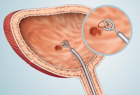
There's no routine test for bladder cancer. But if you're at high risk or have symptoms, your doctor may first order a urine test. If needed, a procedure called cystoscopy lets your doctor see inside the bladder with a slender lighted tube with a camera on the end. The cystoscope can be used to remove small tissue samples (a biopsy) to be examined under a microscope. A biopsy is the best way to diagnose cancer.
Imaging
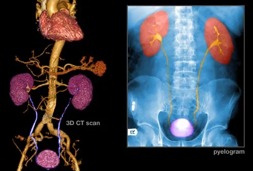
If cancer is found, imaging tests can show whether it has spread beyond the bladder. An intravenous pyelogram uses dye to outline the kidneys, bladder, and ureters, the tubes that carry urine to the bladder. CT and MRI scans give more detailed images of these, and can show the lymph nodes nearby. An ultrasound uses sound waves, instead of radiation, to produce images. Additional imaging tests look for cancer in the lungs and bone.
Types of Bladder Cancer
The main types of bladder cancer are named for the type of cells that become cancerous. The most common is transitional cell carcinoma, which begins in the cells that line the inside of the bladder. Squamous cell carcinoma and adenocarcinoma are much less common.
Stages of Bladder Cancer
Stage 0: Cancer stays in the inner lining.
Stage I: Cancer has spread to the bladder wall.
Stage II: Cancer has reached the muscle of the bladder wall.
Stage III: Cancer has spread to fatty tissue around the bladder.
Stage IV: Cancer has spread to the pelvic or abdominal wall, lymph nodes, or distant sites such as bone, liver, or lungs.
Surgery
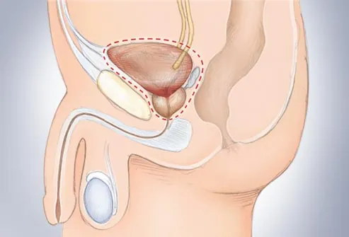
Transurethral surgery is most often done for early-stage cancers. If cancer has invaded more of the bladder, the surgeon will most likely perform either a partial cystectomy, removing a portion of the bladder, or a radical cystectomy, to remove the entire bladder. For men, the prostate and urethra may also be removed. For women, the uterus, fallopian tubes, ovaries, and part of the vagina may also be removed.
After Surgery
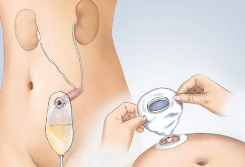
If your entire bladder must be removed, your surgeon will construct another means of storing and passing urine. A piece of your intestine may be used to create a tube that allows urine to flow into an external urostomy bag. In some cases, an internal reservoir -- drained via a catheter -- can be constructed. Newer surgeries offer the possibility of normal urination through the creation of an artificial bladder.
Chemotherapy
Chemotherapy involves drugs designed to kill cancer cells. These drugs may be given before surgery to shrink tumors, making them easier to remove. Chemotherapy is also used to destroy any cancer cells left after surgery and to lower the chances that the cancer will return. Hair loss, nausea, loss of appetite, and fatigue are common side effects. The drugs can be given by vein or directly into the bladder.
Immunotherapy
This type of treatment is delivered directly to your bladder, so it doesn’t treat cancer that has spread beyond it. One treatment, Bacillus Calmette-Guerin therapy, sends in helpful bacteria through a catheter. It triggers your immune system to attack the cancer. Flu-like symptoms are a common side effect of the once-a-week treatment. Immunotherapy may be used after surgery to reduce the risk of recurrence.
Radiation
Radiation uses invisible, high-energy beams, like X-rays, to kill cancer cells and shrink tumors. It's most often given from outside the body by machine. Radiation is often used in tandem with other treatments, such as chemotherapy and surgery. For people who can't undergo surgery, it may be the main treatment. Side effects can include nausea, fatigue, skin irritation, diarrhea, and pain when urinating.
Bladder Cancer Survival Rates
Survival rates are closely tied to the stage at diagnosis. About half of bladder cancers are caught when the disease is confined to the inner lining of the bladder. Nearly 100% of these people will live at least five years, compared to people without bladder cancer. The more advanced the cancer, the lower this figure becomes. But keep in mind that these rates are based on people diagnosed from 1988 to 2001. The treatments and outlook may be better for cancers diagnosed today. And each person’s case is different.
Sex After Bladder Cancer Treatment
Surgery can damage sensitive nerves, making sex more difficult. Some men may have trouble having an erection, though for younger patients, this often improves over time. When the prostate gland and seminal vesicles are removed, semen can no longer be made. Women may also have trouble with orgasm, and may find sex less comfortable. Be sure to discuss treatment options with your doctor.
Living With Bladder Cancer
Cancer is a life-changing experience. And although there's no surefire way of preventing a recurrence, you can take steps to feel and stay healthy. Eating plenty of fruits, veggies, whole grains, and keeping to modest portions of lean meat is a great start. If you smoke, stop. Limit alcohol to one or two drinks a day, if you drink. Daily exercise and regular checkups will also support your health and give you peace of mind.
New and Experimental Treatments
Several new treatments may prove useful in treating bladder cancer. Photodynamic therapy, used in early stage cancers, uses a laser light to activate a chemical that kills cancer cells. Some gene therapies use lab-created viruses to fight cancer. And targeted therapies aim to control the growth of cancer cells. You may be eligible to participate in a clinical trial of these or other cutting-edge treatments.
ps- this is only for information, always consult you physician before having any particular food/ medication/exercise/other remedies.
ps- those interested in recipes are free to view my blog-
http://gseasyrecipes.blogspot.com/
for info about knee replacement, you can view my blog-
http://Knee replacement-stick club.blogspot.com/
for crochet designs
http://My Crochet Creations.blogspot.com
for crochet designs
http://My Crochet Creations.blogspot.com
Labels: adenocarcinoma., bladder cancer, bladder changes, bladder stones, blood, chemotherapy, cystectomy, cystoscope, Immunotherapy, MRI, RADIATION, Squamous Cell Carcinoma(SCC), transitional cell, urine, UTI
posted by G S Iyer at 9:28 AM
0 comments


