Wednesday, September 30, 2026

The 10-Minute Heart Scan That Can Save Your Life

Most of us know our blood pressure. Many of us know our cholesterol numbers. But very few people know what is actually happening inside the arteries that feed their heart. That is a shame, because those arteries can quietly fill with plaque for decades without causing a single symptom.

Today, a quick and painless scan can show doctors how much plaque has built up in your heart arteries, long before it causes chest pain or a heart attack. In March 2026, America's leading heart organizations released new cholesterol guidelines that give this kind of heart imaging a bigger role than ever before. Here is what you need to know about heart artery scans, who should consider one, what the process looks like, and what your results really mean. 

Why Your Heart Arteries Deserve Attention 

Your heart is a muscle, and like any muscle, it needs a steady supply of oxygen-rich blood. That blood arrives through the coronary arteries, a network of vessels that wraps around the outside of the heart. Over the years, cholesterol, fat, calcium, and inflammatory cells can collect in the walls of these arteries, forming what doctors call plaque. This process is known as atherosclerosis, and when it affects the heart arteries, the result is coronary artery disease.

The numbers are sobering. In the United States, coronary heart disease affects more than 20.5 million adults and causes more than 370,000 deaths every year. It is the single largest cause of death among all heart and blood vessel diseases, responsible for about 38% of those deaths. In fact, heart disease and stroke together claimed more American lives in 2023 than all forms of cancer and chronic lung disease combined. 

Roughly every 40 seconds, someone in the United States has a heart attack. That adds up to about 605,000 first heart attacks and another 200,000 repeat attacks each year. The average age of a first heart attack is about 65 for men and 72 for women.

The Problem: Plaque Doesn't Announce Itself 

Here is the most troubling part. Plaque builds up silently. You can feel perfectly healthy, walk your dog every morning, and still have significant buildup in your heart arteries. About half of men and nearly two-thirds of women who die suddenly from coronary heart disease never had any previous symptoms. For many people, the very first sign of heart disease is a heart attack. 

There is another surprise. Many heart attacks do not come from arteries that were already badly narrowed. They often start when a plaque that was only causing a mild or moderate narrowing suddenly cracks open. The body rushes to repair the crack by forming a blood clot, and that clot can block the artery completely within minutes. This is one reason why a traditional treadmill stress test, which looks for arteries that are already narrowed enough to limit blood flow, can come back normal in someone who still has plenty of dangerous plaque. 

That is exactly the gap heart artery imaging is designed to fill. Instead of asking whether blood flow is already limited, it asks a simpler and earlier question: is plaque there at all, and how much?

How Common Is Plaque as We Age? 

Plaque becomes more common with every passing decade. In one of the largest American studies on the subject, which followed thousands of adults between the ages of 45 and 84, about 60% of men and nearly 40% of women already had measurable calcium in their heart arteries. Calcium is a telltale sign of plaque.
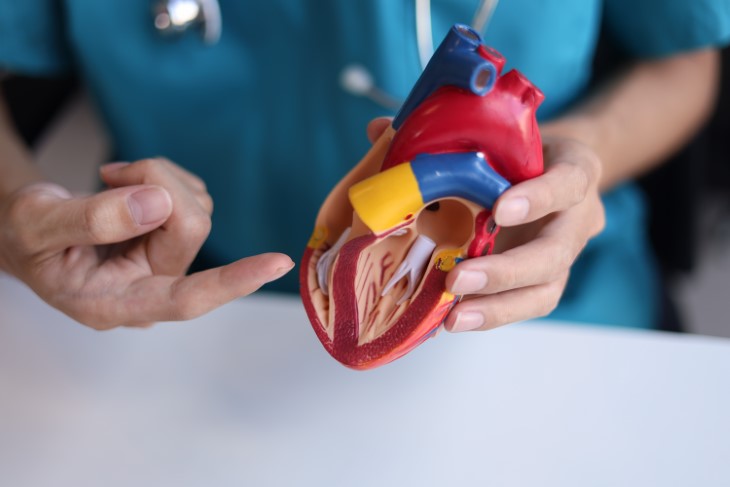

Heart test arteries
By age 75, only about 1 in 9 people have completely clean arteries on a calcium scan, and by age 90, more than 95% of people have at least some calcium. Men tend to develop plaque earlier than women, usually by about 10 years, though women catch up after menopause. 

The good news is that finding plaque early gives you time to act. Plaque can be stabilized, and its growth can be slowed dramatically with the right lifestyle changes and medications. 

The Main Types of Heart Artery Scans

Heart test arteries
When people talk about "mapping" their heart arteries, they usually mean one of three tests. Each has a different purpose. 

1. The coronary calcium scan (calcium score test). This is the most common screening test for people without symptoms. It is a quick, low-dose CT scan that detects hardened, calcified plaque in the heart arteries and turns it into a single number called your calcium score. No needles, no dye, and no special preparation are required.

2. Coronary CT angiography. This is a more detailed CT scan that uses contrast dye injected through a vein in your arm. It creates a detailed three-dimensional picture of the inside of your heart arteries, showing both hard, calcified plaque and soft plaque, which the calcium scan cannot see. It can also estimate how narrow each artery has become. Doctors usually order this test for people who have chest pain or other symptoms, rather than as a general screening tool. 

3. Cardiac catheterization (invasive angiography). This is the traditional gold standard. A thin tube is threaded through an artery in the wrist or groin up to the heart, and dye is injected while X-ray images are taken. It is usually reserved for people with symptoms, abnormal test results, or a suspected heart attack. One advantage is that if a serious blockage is found, a cardiologist can often open it with a balloon and stent during the same procedure. 

Does Scanning Actually Prevent Heart Attacks? 

It is a fair question. A scan by itself does not unclog anything. Its power lies in what happens next. 

A large study in Scotland followed more than 3,500 people who came to cardiology clinics with chest pain. Half received standard care, and half also received coronary CT angiography. After 5 years, the scanned group had about 41% fewer deaths from heart disease and nonfatal heart attacks. After 10 years, the benefit was still there, with serious heart events occurring in about 8.3% of the scanned group compared with 10.3% of the standard care group. 

Interestingly, the scanned group did not end up having more stents or bypass surgeries over the long run. The difference came from prevention. When patients and doctors could actually see the plaque, they were more likely to start cholesterol-lowering medication and other protective treatments, and to stick with them. There is something about seeing your own arteries on a screen that makes the risk feel real. 

Who Should Consider a Calcium Scan? 

A calcium scan is not recommended for everyone, and the new 2026 guidelines make clear that it is not meant for universal screening. It is most useful when you and your doctor are unsure whether you need preventive treatment such as a statin. 

Good candidates are generally people in their 40s, 50s, or 60s who have never had a heart problem but do have risk factors such as: 

Borderline high cholesterol 

 Prediabetes 

Excess weight 

High blood pressure 

A current or past smoking habit 

A parent or sibling who had heart disease at a young age 

 For women, a history of early menopause, preeclampsia, or diabetes during pregnancy 

Inflammatory conditions such as rheumatoid arthritis, lupus, or psoriasis

On the other hand, the test is usually not helpful for people who have already had a heart attack, a stent, or bypass surgery, since their risk is already known to be high. It is also not needed for people with very high LDL cholesterol (190 mg/dL or above), who need treatment regardless, or for people at very low risk, such as most adults under 40. 

What the Process Looks Like 

If you have never had a calcium scan, you may be surprised by how simple it is. The entire appointment usually takes 10 to 15 minutes, and the scan itself takes only a few seconds. 

Before the scan: There is no need to fast. You may be asked to avoid caffeine and smoking for a few hours beforehand, since both can speed up your heart rate and blur the images. Wear comfortable clothing without metal, or expect to change into a gown.

During the scan: A technician places a few small sticky pads on your chest to track your heartbeat, so the machine can take pictures between beats when the heart is still. You lie on your back on a table that slides into the open, ring-shaped CT scanner. You will be asked to hold your breath for several seconds while the images are taken. There are no needles and no dye. 

After the scan: You can go straight back to your normal day. A radiologist or cardiologist reviews the images, and your doctor typically receives the results within a few days. 

Radiation: The dose is low, roughly 1 to 2 millisieverts, which is similar to a mammogram and equal to a few months of the natural background radiation we all absorb from the environment. 

Cost: Because it is considered a screening test, most insurance plans, including Medicare, usually do not cover a calcium scan. The good news is that it is relatively affordable, typically between $75 and $300 out of pocket, and many imaging centers offer a flat cash price. Some centers accept self-referrals, but it is best to go through your doctor so the results are interpreted in context. 

Coronary CT angiography is a slightly longer process. You will have an IV placed for the contrast dye, and you may be given medication to slow your heart rate for clearer images. The radiation dose is somewhat higher, and people with kidney problems or a dye allergy need special precautions. When ordered for symptoms, it is usually covered by insurance. 

What Your Calcium Score Means 

Your result will be a number, usually called an Agatston score after the doctor who developed the method. Your report may also include a percentile, which compares your score with other people of the same age, sex, and background. Under the 2026 guidelines, each score range is now linked to specific cholesterol goals.

Score of 0: No calcified plaque was found. Your risk of a heart attack over the next several years is very low. Many people in this group can safely hold off on statins, unless they smoke, have diabetes, or have a strong family history of early heart disease. The guidelines suggest considering a repeat scan in 3 to 7 years. 

Score of 1 to 99: Mild plaque is present. Heart disease has officially begun, even if it is early. Doctors generally aim to bring LDL cholesterol below 100 mg/dL, often with a statin and lifestyle changes. 

Score of 100 to 299: Moderate plaque. Your risk is meaningfully higher. The goal is to cut LDL cholesterol by at least half and bring it below 70 mg/dL. 

Score of 300 or higher: Extensive plaque and a high risk of a heart attack. The target LDL is below 55 mg/dL, which often requires a combination of medications.

Score above 1,000: Very extensive plaque. People in this range are treated as aggressively as those who have already had a heart attack. 

One important point: a high calcium score does not automatically mean you have a blocked artery or need a stent. It measures how much plaque you have overall, not how narrow any single artery is. If you have no symptoms, the main response to a high score is usually medication and lifestyle changes, not surgery. If you do have symptoms, such as chest pressure or unusual shortness of breath during activity, your doctor may order further testing. 

A Bonus Many People Miss 

If you have ever had a regular chest CT scan, for example for lung cancer screening or to check a cough, the radiologist may have noticed calcium in your heart arteries. Under the new guidelines, calcium spotted on an ordinary chest CT can now be used to guide treatment decisions. If you have had such a scan, it is worth asking your doctor whether any coronary calcium was mentioned in the report. Keep in mind that the reverse is not true: a regular chest CT that shows no calcium is not a substitute for a proper calcium scan. 

The Limitations Worth Knowing 

No test is perfect, and a calcium scan has a few blind spots. It only detects calcified plaque, so it can miss younger, softer plaque, which is sometimes the more unstable kind. That is why a score of zero is reassuring but not a lifetime guarantee, especially for smokers and people with diabetes. 

The scan can also pick up incidental findings in the lungs or other nearby structures, such as small nodules. Most turn out to be harmless, but they can lead to follow-up scans and some worry. It is a good idea to discuss this possibility with your doctor beforehand. 

Finally, a calcium scan does not replace the basics. Blood pressure, cholesterol, blood sugar, and a simple one-time blood test for lipoprotein(a), an inherited type of cholesterol particle, all remain important pieces of the puzzle. 

What to Do After You Get Your Results 

Whatever your score, the steps that protect your arteries are the same, and they work at any age:Stop smoking, which is the single most powerful step for your arteries: Aim for about 150 minutes of moderate activity a week, such as brisk walking Eat plenty of vegetables, fruit, whole grains, beans, nuts, and fish, and limit processed meats and sugary foods Keep your blood pressure and blood sugar under control Get 7 to 9 hours of sleep a night Take prescribed medications consistently, even when you feel fine.


This is only for your information, kindly take the advice of your doctor for food, medicines, exercises and so on.   


Labels: , , , , , , , , , , , ,

Wednesday, July 29, 2026

What Causes Those Bumps on Our Scalp?

When you think about the health of your scalp, most of the time you're probably focused on your hair. However, it turns out there's a lot going on with the skin underneath your hair. So, in addition to frizziness or hair loss, you could be facing dandruff or bumps on your scalp, too. The good news is that if your scalp is healthy, you'll have a better chance of growing healthy hair, according to evidence from the National Institutes of Health. When the scalp is unhealthy, it can do the opposite. When something in your scalp goes awry, it can lead to uncomfortable rashes, bumps, flakes, sores, scabs and even hair loss.

The Anatomy of the Scalp 

The skin on the scalp is different than that on the rest of the body: Besides the obvious profusion of hair follicles, the scalp has more sebaceous glands — tiny glands found in hair follicles (and some hairless parts of the body as well) that release sebum, an oily substance that acts as a lubricant for hair and skin. All of this makes the scalp a particularly rich environment for microbes, which can bring on various problems — including some that require medical attention.

If you're dealing with itchy, unsightly or just plain annoying scalp issues, use the info below to help figure out if one of these common scalp conditions could be the root cause. Once you know what's going on, you can better treat the underlying condition. 

Since it's nearly impossible to get a good look at the top or back of your own head, it's a great idea to have a board-certified dermatologist examine your scalp and come up with a treatment plan to help you find relief. 

Head Lice 

These tiny insects live in the hair on people's heads (including eyebrows and eyelashes!) and feed on human blood. While they don't spread any diseases and they can't jump or fly, lice can crawl from one person to another if they touch, according to the Centers for Disease Control and Prevention (CDC). When a person is allergic to a lice bite, it can itch.

What it looks like: 

Lice eggs (nits) are usually attached to the hair shaft and may be confused with scabs or dandruff flakes. Adult lice are about the size of a sesame seed and tan or gray in color; they may appear darker on darker hair. There may be sores on the scalp if the person has been scratching. 

The causes: Usually lice are spread when one person's head touches another person's. Young kids are the biggest culprits, but it's important to note that a lice infestation does not mean a person is dirty. It's also possible — but not likely — to get lice from shared items like clothes, pillows or combs.

The treatments: Head lice can usually be taken care of with an OTC shampoo treatment and a lice comb. If that doesn't work, a doctor can prescribe additional medications. You should also vacuum, wash the infected person's clothes and bedding and disinfect their combs and brushes, according to the CDC. 

Lichen Planus 

Lichen planus (LP) is an inflammatory disorder that affects the skin and mucus membranes. Research shows it's common and can show up nearly anywhere on the body. This includes the mouth, genitals, nails, legs and the scalp (just to name a few spots!).

What it looks like: 

LP usually appears as small purple or red irritated bumps on the scalp and in some cases, it can cause hair loss. On other parts of the body, it may manifest in a different way, according to the American Academy of Dermatology (AAD). 

The causes: No one is exactly sure what causes a person to develop LP, but the AAD says these are some likely triggers: 

1. In rare instances, metal dental fillings have been linked with LP. 

2. People with Hepatitis C are more likely to have LP. 

3. Certain medications seem to spark the development of LP so tell your dermatologist about everything that you take. 

4. Genetics may also come into play — one particular type of LP called bullous lichen planus tends to run in families. 

The treatments:  

1.Topicals like corticosteroid, retinoic acid, tacrolimus ointment and pimecrolimus cream can be applied to skin where there are LP lesions. 

2. Antihistamines and retinoic acid can be taken in pill form. 

3. Using PUVA therapy, patients either soak parts of their skin in water that contains psoralens (plant compounds that make skin more sensitive to UVA rays) or they take psoralens orally. Then, affected skin is exposed to ultraviolet A (UVA) light.

Seborrheic Dermatitis (Dandruff)

Seb derm, as most dermatologists call it, is one of the most common scalp conditions. In babies, it's called cradle cap; in adults, it causes red, itchy patches with greasy scales or flakes. People usually refer to it as dandruff when it's on the scalp, but seb derm when it affects the face, nose, eyebrows, ears or chest. Research estimates that half of all adults in the world have dandruff so if you're struggling with it you're definitely not alone. Seb derm often comes and goes with triggers such as stress and seasonal changes — it is usually worse in winter and early spring. It's often best to treat seborrheic dermatitis early and aggressively to avoid complications such as hair loss.

What it looks like: 

Greasy scales and dandruff flakes 

The causes:

1. A yeast (fungus) called malassezia, which is in the oil secretion on the skin, is thought to be the culprit, though doctors don't know for sure. 

2. Parkinson's disease, depression, diabetes, a weakened immune system and obesity may make a person more likely to have seb derm. So can recovering from a stressful health event, like a heart attack. 

3. Dry skin can result in flakes as can a sensitivity to certain hair products so it's helpful to look at your scalp care regimen to see if something other than a fungus is causing your flakes. 

The treatments: 

1. A prescription strength anti-yeast shampoo called ketoconazole can help. Since it can be very drying to the hair, applying the shampoo directly to your scalp 5 to 10 minutes before wetting the hair and then lathering only the scalp area before you rinse it off is recommended. Following with a moisturizing shampoo and conditioner on the hair shaft can prevent the dryness that prescription shampoos can often cause, especially in more coily hair types. 

2. OTC shampoos or lotions that contain zinc pyrithione, ketoconazole or sulfur may also help. 

3. A topical steroid may be prescribed if the shampoo doesn't work, or in combination with the shampoo if the scales are very dense. 

Psoriasis 

 psoriassis

This condition affects about 1 to 2% of the general population and anyone can get it, at any age. Psoriasis can appear on any part of the body, but according to the National Psoriasis Foundation, as many as 45% to 56% of those with psoriasis have it on their scalp. And that kind is one of the most difficult types to treat. One way to distinguish it from dandruff: The buildup of skin cells creates scaly patches that are dry, rather than oily. And those patches can itch, hurt, crust up and bleed.

What it looks like:
Pink plaques with a silvery scale. On darker skin tones, the plaques may be dark red or purplish. The causes: 1. Psoriasis is thought to be an immune dysregulation around skin cells that causes the cells in the top layer of skin to go into overdrive. 2. Stress can make the situation worse, because it causes an increase in the hormone cortisol, which in turn can boost inflammation.

The treatments:
 

1. Topical steroids, which control the inflammatory response, are the most common treatment. 

2. Injections called biologics that help suppress what's causing the flare-ups in tougher cases. 

3. An excimer laser can work on individual spots that haven't responded to other therapies. 

4. A gentler hair routine can also help. Not shampooing your hair every day may lessen the number of skin rashes, or a therapeutic oil cream can do wonders. 

Folliculitis  scalp skin conditions

Folliculitis, a common skin condition, is basically inflammation of the hair follicle; it appears as little pus bumps scattered throughout the scalp that look similar to acne and can spread. The bumps can sometimes feel sore.

What it looks like: 

 Scattered pus bumps. On darker skin tones, the bumps may be a dark shade of red or light purple around the periphery. 

The causes:  

1. Bacteria (most commonly a staph infection) or a fungal infection. 

2. Irritation from shaving, heavy conditioners, potent hair dyes and excessive sweating. 

The treatments: 

1. Topical or oral antibiotics may be prescribed if your doctor determines via a culture that bacteria is causing your scalp woes.

2. Anti-inflammatory medications, such as topical steroid formulations, can be effective if there isn't an infectious agent present. 

Hair Loss 

Hair loss can be due to a number of different factors, including genetics, medications, inflammation, hormonal abnormalities and hair fragility. Regardless of the cause of hair loss, it can be extremely distressing. If you notice your hair breaking or shedding, or you see bald spots on your scalp, make an appointment to get evaluated by a health care provider immediately.

What it looks like: Anything from hair breaking or shedding, to bald spots on your scalp. 

The causes: 

1. Anemia, vitamin deficiency, thyroid abnormalities, irregular hormone levels (especially after pregnancy) and autoimmune markers can all lead to hair loss. 

2. Extreme stress, such as the death of a loved one, can have an impact on your whole body including your hair. 

3. Major weight loss (sometimes related to a high-protein diet) can also trigger hair loss. 

4. Infection and illness can cause a massive shedding months after the incident — a condition called telogen effluvium. For instance, a survey conducted by Survivor Corps found that nearly 68% of 1,700 COVID-19 survivors that were interviewed reported experiencing hair loss in the wake of their coronavirus infection. 

Age can also play a role: As estrogen levels decrease between childbearing age and menopause, our hair may thin out. Years of bleaching, dyeing, straightening, perming and rocking tight, slicked-back hairstyles can also catch up to us.

The treatments:
 

1. Rogaine, a solution that thickens existing hair, and spironolactone, a pill that helps block hormones called androgens, can be helpful in cases of hormonal thinning. 

2. Eating a well-balanced diet is also useful when experiencing hair health woes, and talking to your doctor about whether you're deficient in iron or specific vitamins. 

3. Taking a break from over-processing your hair and wearing more natural styles is recommended. 

4. Platelet-rich plasma (PRP) is an effective option for androgenetic alopecia (also called male and female pattern hair loss) and the residual thinning after telogen effluvium. During PRP, a patient's blood is drawn and the plasma, which is rich in various growth factors effective for stimulating hair regrowth, is isolated and injected or applied to the scalp with microneedling.

Cysts 

Cysts can occur on the skin, just like they can show up on other organs. The good news: Scalp cysts are common and easy to identify. They are usually bumps the size of marbles or grapes, and you can feel them when your comb or brush runs over them. If you're unsure about a bump or you can't get a good look at your scalp, that's a smart time to consult a dermatologist.

What it looks like:
A skin-colored or pink-red smooth bump

 The cause: For the most part, scalp cysts are mostly genetic. 

The treatments: 

1. Leave them alone. This is definitely an easy option if the scalp cysts aren't bothering you, since they are mostly benign and shouldn't raise any concern. 

2. Have them drained: In some cases, however, scalp cysts can get infected or irritated, which causes pain. Incision and drainage can help calm the symptoms, but won't remove the cyst in its entirety. 

3. Consider antibiotics: These can also help relieve symptoms of an inflamed or infected cyst. 

4. Have them surgically removed by a medical professional. Cysts contain a fluid-filled sac. Unless the sac is removed, the cyst can recur, thus surgery is the only effective option to completely remove a cyst and make sure it doesn't recur. But the surgical removal is very simple and done with just a local anesthetic. While it does leave a scar, it's often not noticeable on the scalp since it's hidden by hair.

Ringworm 

scalp skin conditions 

Also called tinea capitis, ringworm is a contagious fungal infection that usually happens in little kids, but occasionally infects adults too. Despite the name, no worm is involved: The condition got its name from its circular shape. The rash is usually painful and scaly and sometimes causes enlarged lymph nodes. It also can lead to bald spots, broken hairs and black dots on the skin. 

What it looks like: Scaly rash and bald spots

The cause: According to the Mayo Clinic, ringworm can be spread human to human, animal to human (cats are a common source) and object (like clothing, towels, bedding, combs and brushes) to human. Ringworm can also be transferred by indirect contact through a swimming pool or hot tub. 

The treatment: Antifungal medication taken daily for 4 to 6 weeks usually eliminates ringworm. Ringworm is no longer contagious 48 hours after starting treatment. Until then, you want to avoid contact with others, as tinea capitis can spread through contact with other areas of the body and other people.

Allergic Contact Dermatitis (ACD)

 scalp skin conditions

This rash is a reaction to the scalp coming into direct contact with an allergen that results in a response from the immune system — sometimes days after the contact, so it can be difficult to determine what the body is reacting to. ACD can be painful, itchy and even oozing in some cases. The symptoms of a severe case can often be mistaken for infection. What it looks like: Itchy redness. On darker skin tones, it may look pink, grayish or purple. 

The causes:  

1. Paraphenylenediamine (PPD) is a chemical that is widely used as a permanent hair dye and it can wreak havoc on the skin and is one of the most common causes of scalp allergic contact dermatitis. 

2. The chemicals/fragrances in shampoos and conditioners are also a common cause of ACD. 

3. Other relevant possible allergens include propylene glycol, Cocamidopropyl betaine, methylisothiazolinone (MI) or a combined formulation of methylchloroisothiazolinone and MI. 

 The treatments: 

1. Identifying the trigger and eliminating it. As stated, that can be difficult to determine; sometimes a patch test is needed to figure out what the body is allergic to. 

2. Topical steroid gel, foam, cream or ointment: Two to three weeks of treatment should help decrease inflammation and clear up your skin. 

3. Systemic steroids may be prescribed in severe cases - consult your doctor.

In Conclusion... 

Understanding the various conditions that can affect your scalp is an important step toward maintaining both scalp and hair health. From common issues like dandruff and folliculitis to more complex conditions such as psoriasis and lichen planus, each scalp condition has distinct characteristics, causes, and treatment options. The key to effective management lies in proper identification and timely intervention. 

While some scalp conditions can be treated with over-the-counter products and lifestyle modifications, others require professional medical attention and prescription treatments. If you're experiencing persistent itching, unusual bumps, significant flaking, hair loss, or any other concerning symptoms, don't hesitate to consult a board-certified dermatologist. Early diagnosis and treatment can prevent complications and often lead to better outcomes. 

Remember that a healthy scalp is the foundation for healthy hair growth. Simple practices like maintaining good hygiene, avoiding harsh chemicals when possible, managing stress levels, and being mindful of potential allergens in your hair care products can go a long way in preventing many scalp issues. Additionally, if you notice any changes in your scalp health or if symptoms persist despite treatment, seeking professional evaluation is always the wisest course of action. 

Your scalp deserves the same attention and care as the rest of your skin. By staying informed about these common conditions and their treatments, you're better equipped to recognize problems early and take appropriate action. Whether dealing with a minor irritation or a more serious condition, proper care and medical guidance can help restore your scalp to optimal health.


This is only for your information, kindly take the advice of your doctor for food, medicines, exercises and so on.   


Labels: , , , , , , , , , , , , , ,