Wednesday, February 05, 2020

Scientists Grow 'Yarn' Out of Human Skin Cells So They Can Literally Stitch People Up

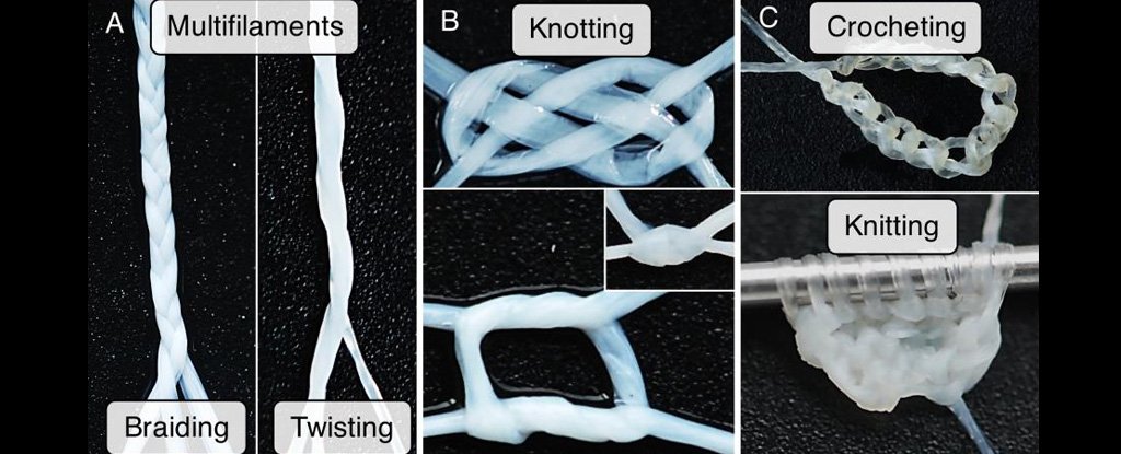
A team of researchers at the French National Institute of Health and Medical Research in Bordeaux have grown yarn from human skin cells that they call a "human textile" - and they say it could be used by surgeons to close wounds or assemble implantable skin grafts.
main article image

"These human textiles offer a unique level of biocompatibility and represent a new generation of completely biological tissue-engineered products," the researchers wrote in a paper published in the journal Acta Biomaterialia.

The key advantage of the gruesome yarn is that unlike conventional synthetic surgical materials, the material doesn't trigger an immune response that can complicate the healing process, according to New Scientist.

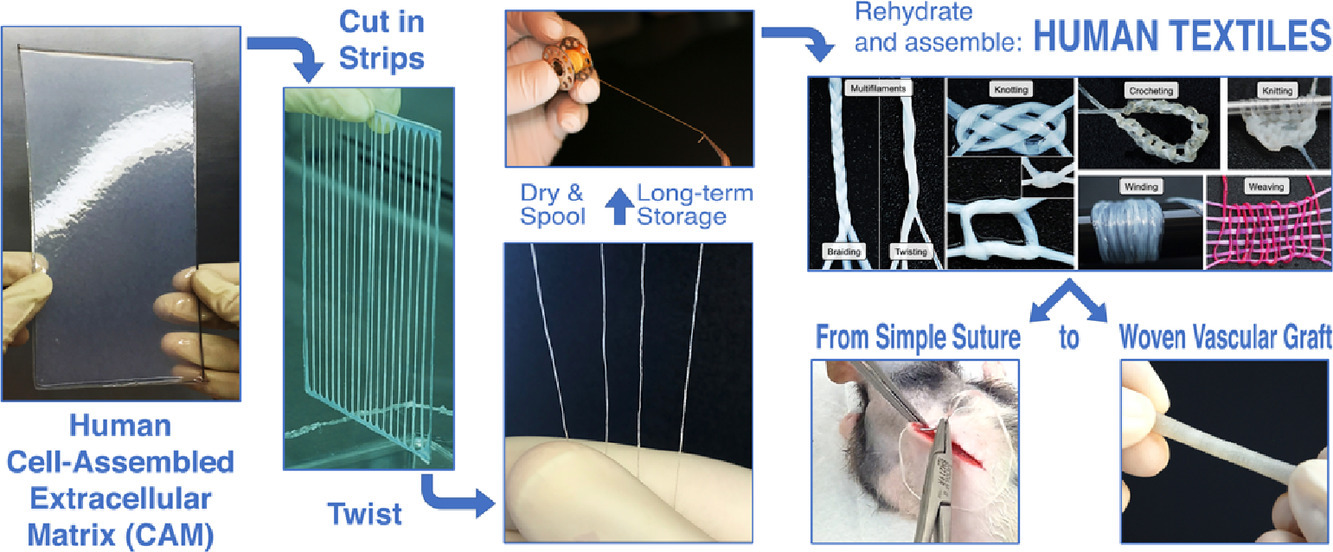
To create it, according to the magazine, the researchers cut sheets of human skin cells into long strips - and then "wove" them into a yarn-like material that can be fabricated into a variety of shapes.

"We can sew pouches, create tubes, valves and perforated membranes," lead researcher Nicholas L'Heureux told New Scientist.

"With the yarn, any textile approach is feasible: knitting, braiding, weaving, even crocheting."
graphical abstract human skin yarn


So far, the researchers have used the special yarn to stitch a rat's wounds and help it fully heal over two weeks. They even created a skin graft, using a custom-made loom, to seal a sheep's artery and stop it from leaking.

The work builds on prior research by the same team in which they produced sheets of biomaterial and rolled them into artificial blood vessels.

This article was originally published by Futurism. Read the original article.

This is only for your information, kindly take the advice of your doctor for medicines, exercises and so on.     
https://gscrochetdesigns.blogspot.com. one can see my crochet creations  
https://gseasyrecipes.blogspot.com. feel free to view for easy, simple and healthy recipes    
https://kneereplacement-stickclub.blogspot.com. for info on knee replacement
 

Labels: , , , , ,

Saturday, January 18, 2020

Ingestible medical devices can be broken down with light

A variety of medical devices can be inserted into the gastrointestinal tract to treat, diagnose, or monitor GI disorders. Many of these have to be removed by endoscopic surgery once their job is done. However, MIT engineers have now come up with a way to trigger such devices to break down inside the body when they are exposed to light from an ingestible LED.

The new approach is based on a light-sensitive hydrogel that the researchers designed. Incorporating this material into medical devices could avoid many endoscopic procedures and would give doctors a faster and easier way to remove devices when they are no longer needed or are not functioning properly, the researchers say. 

"We are developing a set of systems that can reside in the gastrointestinal tract, and as part of that, we're looking to develop different ways in which we can trigger the disassembly of devices in the GI tract without the requirement for a major procedure," says Giovanni Traverso, an assistant professor of mechanical engineering, a gastroenterologist at Brigham and Women's Hospital, and the senior author of the study.

In a study in pigs, the researchers showed that devices made with this light-sensitive hydrogel can be triggered to break down after being exposed to blue or ultraviolet light from a small LED. 

Ritu Raman, a postdoc at MIT's Koch Institute for Integrative Cancer Research, is the lead author of the paper, which appears today in Science Advances. Other authors of the paper are former technical associates Tiffany Hua, Jianlin Zhou, Tina Esfandiary, and Vance Soares; technical associates Declan Gwynne, Joy Collins, and Siddartha Tamang; graduate student Simo Pajovic; Division of Comparative Medicine veterinarian Alison Hayward; and David H. Koch Institute Professor Robert Langer.

Controlled breakdown
Over the past several years, Traverso and Langer have developed many ingestible devices designed to remain in the GI tract for extended periods of time. They have also worked on a variety of strategies to control the breakdown of such devices, including methods based on changes in pH or temperature, or exposure to certain chemicals. 

"Given our interests in developing systems that can reside for prolonged periods in the gastrointestinal tract, we continue to investigate a range of approaches to facilitate the removal of these systems in the setting of adverse reaction or when they are no longer needed," Traverso says. "We're really looking at different triggers and how they perform, and whether we can apply them to different settings."

In this study, the researchers explored a light-based trigger, which they believed could offer some advantages over their earlier approaches. One potential advantage is that light can act at a distance and doesn't need to come into direct contact with the material being broken down. Also, light normally does not penetrate the GI tract, so there is no chance of accidental triggering. 

To create the new material, Raman designed a light-sensitive hydrogel based on a material developed in the lab of Kristi Anseth, a former Langer lab postdoc who is now a professor of chemical and biological engineering at the University of Colorado at Boulder. This polymer gel includes a chemical bond that is broken when exposed to a wavelength of light between 405 and 365 nanometers (blue to ultraviolet). 

Raman decided that instead of making a material composed exclusively of that light-sensitive polymer, she would use it to link together stronger components such as polyacrylamide. This makes the overall material more durable but still allows it to break apart or weaken when exposed to the right wavelength of light. She also constructed the material as a "double network," in which one polymer network surrounds another.

"You're forming one polymer network and then forming another polymer network around it, so it's really entangled. That makes it very tough and stretchy," Raman says.

The material's properties can be tuned by varying the composition of the gel. When the light-sensitive linker makes up a higher percentage of the material, it breaks down faster in response to light but is also mechanically weaker. The researchers can also control how long it takes to break down the material by using different wavelengths of light. Blue light works more slowly but poses less risk to cells that are sensitive to damage from ultraviolet light.

Deflated by light
The gel and its breakdown products are biocompatible, and the gel can be easily molded into a variety of shapes. In this study, the researchers used it to demonstrate two possible applications: a seal for a bariatric balloon and an esophageal stent. Standard bariatric balloons, which are sometimes used to help treat obesity, are inflated in a patient's stomach and filled with saline. After about six months, the balloon is removed by endoscopic surgery.

In contrast, the bariatric balloon that the MIT team designed can be deflated by exposing the seal to a tiny LED light, which would in principle be swallowed and then pass out of the body. Their balloon is made of latex and filled with sodium polyacrylate, which absorbs water. In this study, the researchers tested the balloons in pigs and found that the balloons swelled up as soon as they were placed in the stomach. When a small, ingestible LED emitting blue light was placed in the stomach for about six hours, the balloons slowly deflated. With a higher-power light, the material broke down within 30 minutes.

The researchers also molded the light-sensitive gel into an esophageal stent. Such stents are sometimes used to help treat esophageal cancer or other disorders that cause a narrowing of the esophagus. A light-triggerable version could be broken down and passed through the digestive tract when no longer needed. 

In addition to those two applications, this approach could be used to create other kinds of degradable devices, such as vehicles for delivering drugs to the gastrointestinal tract, according to the researchers. 

"This study is a proof of concept that we can create this kind of material, and now we're thinking about what are the best applications for it," Traverso says.

This is only for your information, kindly take the advice of your doctor for medicines, exercises and so on.     
https://gscrochetdesigns.blogspot.com. one can see my crochet creations  
https://gseasyrecipes.blogspot.com. feel free to view for easy, simple and healthy recipes    
https://kneereplacement-stickclub.blogspot.com. for info on knee replacement
 

Labels: , , , , , , , , , , , , ,

Wednesday, January 15, 2020

How urine tests could detect organ transplant rejection and eliminate needle biopsies

Researchers from Georgia Institute of Technology have developed a new screening test with sensors and a urine test to detect transplanted organ rejection without the need for a biopsy needle.

There were approximately 36,528 organ transplants in 2018, according the U.S. Department of Health and Human Services. Transplant rejection happens when a recipient’s immune system attacks the transplanted organ or tissue. Traditionally, the only way to determine if there has been serious damage to a transplanted organ because of rejection is through a biopsy.

The new method’s particles are able to send a fluorescent signal to the urine of a transplant recipient. The researchers have designed the sensor using highly biocompatible components and tested the method in mouse models.

A nanoparticle sensor in the detection method is able to detect a T cell weapon called granzyme B, which turns the transplanted organ’s cells against itself in a process known as apoptosis.

“Before any organ damage can happen, T cells have to produce granzyme B, which is why this is an early detection method,” Gabe Kwong, a researcher on the study, said in a press release.

The nanoparticles are crafted together in the middle like a ball using iron oxide. They are double-coated with dextran and polyethylene glycol to help keep the body from getting rid of it too quickly.

“This is sensitive enough to possibly detect budding rejection before you see significant injury to the transplanted organ and that could help clinicians treat early to prevent damage,” Dr. Andrew Adams, a researcher on the study and an associate professor of surgery at Emory University School of Medicine. “Right now, most tests are aimed at organ dysfunction, and sometimes they don’t signal there is a problem until organ function is below 50%.”

Amino acid bristles stick out from the iron ball with fluorescent reporter molecules attached at the tip.

The particles are then injected intravenously. They are small enough to accumulate in tissues of struggling transplanted organs to watch for rejection, according to the researchers. The particles are too big to accumulate in native tissue or to pass through kidneys and out of the body.

Once the T cells start making granzyme B, amino acid strands in the organ’s cells are cut off and the cells can begin to die.

“The nanoparticles’ bristles mimic granzyme’s amino acid targets in the cells, so the enzyme cuts the bristles on the nanoparticle at the same time,” Kwong said. “That releases the reporter molecules, which are so small that they easily make it through the kidney’s filtration and go into the urine.”

In animal tests, the urine glows and was able to be seen in bladders in near-infrared images.

The researchers now plan to use the sensor to detect other causes of transplant rejection like attacks by antibodies.

“Antibodies kill their target cells through similar types of enzymes. In the future, we envision a single sensor to detect both types of rejection,” said Kwong.

Kwong and the researchers tested the urine test mouthed in small skin grafts on mice and were able to get a very clear timely signal from the nanoparticle sensor.

“This method could be adapted to tease out multiple problems like rejection, infection or injury to the transplanted organ,” Adams said. “The treatments for all of those are different, so we could select the proper treatment or combination of treatments and also use the test to measure how effective treatment is.”

Biopsies are the current method for detecting if an organ transplant is working. However, biopsies can go wrong and the needle can damage tissue.

“This biggest risk of a biopsy is bleeding and injury to the transplanted organ,” Adams said. “Then there’s the possibility of infection. You’re also just taking a tiny fraction of the transplanted organ to determine what’s going on with the whole organ, and you may miss rejection or misdiagnose it because the needle didn’t hit the right spot.”

The researchers suggest that a urine test could have a more global reading on an entire organ while offering other advantages over biopsies.

“The biopsy is not predictive. It’s a static snapshot. It’s like looking at a photo of people in mid-jump. You don’t know if they’re on their way up or on their way down. With a biopsy, you don’t know whether the rejection is progressing or regressing,” Kwong said. “Our method measure biological activity rates, and tells us where things are going.”

Measuring biological activity could also allow clinicians to dose powerful immunosuppressant medications that most transplant patients receive.

“Adjusting the dose is very difficult but very important because heavy immunosuppression increases occurrence of infections and patients who receive it also get cancer more often,” Kwong said.

The research was published in the journal Nature Biomedical Engineering and was funded by the National Institutes of Health, the National Science Foundation and the Burroughs Wellcome Fund.


this is only for your information, kindly take the advice of your doctor for medicines, exercises and so on.     
https://gscrochetdesigns.blogspot.com. one can see my crochet creations  
https://gseasyrecipes.blogspot.com. feel free to view for easy, simple and healthy recipes    
https://kneereplacement-stickclub.blogspot.com. for info on knee replacement
 

Labels: , , , , , , , , , , ,

Monday, January 14, 2019

New biodegradable nanofibre bandage enables faster healing

An international team of scientists has developed a new bandage with a strong antibacterial effect and which can be kept on over long periods of time without needing to be changed, thereby accelerating skin regrowth and reducing the risk of infection. The new dressing material has great potential for future application in the healing of wounds, does not require changing, and can literally be put on over top of one another as they degrade, the Sputnik news agency reported.

The traditional use of an antiseptic in the treatment of wounds, consisting of washing the affected area with a solution and using gauze dressing to increase the outflow of fluids, repeated several times.
However, such treatment, particularly the use of antibiotics kill not only dangerous bacteria but also the useful bacteria. And when dressings are changed, the fragile area undergoing healing is disturbed, causing severe pain to patients.

But according to scientists, the new biocompatible dressing material can act locally on the area of inflammation. As the material slowly releases their antibiotic, the dressings gradually dissolve on the skin.

The material was made using polycaprolactone nanofibres -- a biocompactable, bioresabsorable material -- and attached gentamicin, a broad-spectrum antibiotic, to the fibres.

"What we found was that the action was prolonged: we observed a significant decrease in the number of bacteria even 48 hours after the material was applied," a researcher was quoted as saying.

"Usually, surfaces with an antibacterial effect fulfill their potential within the first day or even the first hours of use," the researcher  explained. In the study, reported in the Materials and Design academic journal, the team performed experiments using three strains of E. coli bacteria, with the strains showing varying levels of resistance to the antibiotics, but nonetheless combated against effectively by the dressing.

The new material is potentially applicable not only for treatment to heal skin, but to treat inflammatory bone diseases such as osteoporosis and osteomyelitis, the report noted.

THIS IS ONLY FOR INFORMATION, ALWAYS CONSULT YOU PHYSICIAN BEFORE HAVING ANY PARTICULAR FOOD/ MEDICATION/EXERCISE/OTHER REMEDIES.                                                                                
 PS- THOSE INTERESTED IN RECIPES ARE FREE TO  VIEW MY BLOG-                                                                                           https://gseasyrecipes.blogspot.com/                                                                                                                                                                             FOR INFO ABOUT KNEE REPLACEMENT, YOU CAN VIEW MY BLOG-                                               
     https:// kneereplacement-stickclub.blogspot.com/                                                                                     
  FOR CROCHET DESIGNS                                                                                                                                                                                                  https://gscrochetdesigns.blogspot.com
  

Labels: , , , , , , , , , ,