Thursday, October 13, 2022

Researchers develop new 3D technique: Innovation to treat Diabetic Foot Ulcer

Diabetes, a lifelong condition that causes a person's blood sugar level to become too high, is among the top ten causes of deaths worldwide. Diabetic foot ulcer (DFU), is a serious complication of diabetes, affecting approximately 25% of diabetic patients. When identified, over 50 per cent are already infected and over 70 per cent of cases result in lower limb amputation. Queen's University Belfast has come out with a new bandage treatment, known as a scaffold, to treat diabetic foot ulcers & also cost-effective while improving patient outcomes.

The 3D printing method was first demonstrated in the 1980s, and since then, the field of 3D printing (3DP) and bioprinting has grown rapidly in recent years due to technological developments and a greater understanding of the capabilities using these technologies, particularly in the development of scaffolds for tissue engineering applications. In the fabrication of 3D (bio) printed scaffolds, the user is firstly required to design a scaffold using computer-aided design (CAD).

Specialist software can then be used to slice the design prior to printing, providing a set of printing instructions for the printer; each 3DP manufacturer provides the software. A wide range of advantages is offered by 3DP, including the capability to rapidly produce elaborate (scaffold) designs of various sizes at low cost. Moreover, 3D-bioprinted scaffolds have shown the ability to achieve high cell loading and maintain high levels of cell viability while also providing structural support to the wound. 

The treatment strategy required for the effective healing of diabetic foot ulcer (DFU) is a complex process that is requiring several combined therapeutic approaches. As a result, there is a significant clinical and economic burden associated in treating DFU. Furthermore, these treatments are often unsuccessful, commonly resulting in lower-limb amputation. 

Graphical abstract
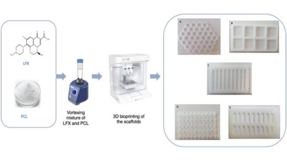
file photo

This new research demonstrates outcomes with significant implications for patient quality of life, as well as decreasing the costs and clinical burden in treating DFU. Recent research has focused on drug-loaded scaffolds to treat DFU. The scaffold structure is a novel carrier for cell and drug delivery that enhances wound healing.

Springler Link Research 

The research, published in Springer Link, was presented by Professor Lamprou at the Controlled Release Society (CRS) Workshop Italy.

Professor Dimitrios Lamprou, a Professor of Biofabrication and Advanced Manufacturing at Queen's School of Pharmacy and corresponding author, explains: "These scaffolds are like windows that enable doctors to monitor the healing constantly. This avoids needing to remove them constantly, which can provoke infection and delay the healing process.
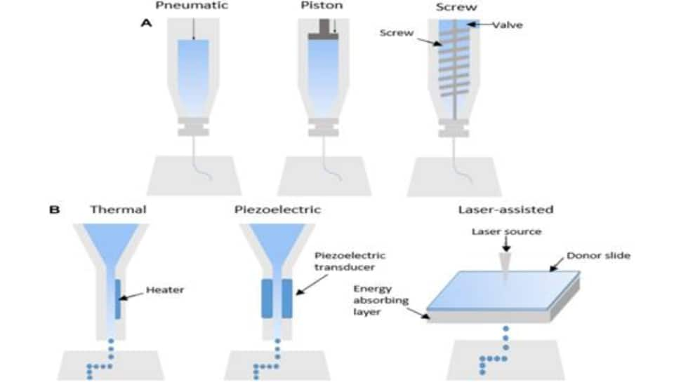
file photo

Schematics showing (a) extrusion-based, (b) inkjet-based, and (c) laser-assisted bioprinting technologies

"The 'frame' has an antibiotic that helps to 'kill' the bacteria infection, and the 'glass' that can be prepared by collagen/sodium alginate can contain a growth factor to encourage cell growth. The scaffold has two molecular layers that both play an important role in healing the wound."

What experts say

According to expert Ashok Jhingan , Sr Phycisian & Sr director BLK Max, "the annual incidence of diabetic foot ulcer worldwide is between 9.1 to 26.1 million. Around 15 to 25% of patients with diabetes mellitus will develop a diabetic foot ulcer during their lifetime.

The majority of patients with diabetes who develop foot ulcers are male (more than two-thirds). Furthermore, most of them are between 40 and 70 years of age. Older patients face significantly more complications than younger ones.

Diabetic peripheral neuropathy is a precipitating factor in almost 90% of diabetic foot ulcers. Chronically high glucose (blood sugar) levels damage nerves, including the sensory, motor and autonomic nerves. Diabetic neuropathy also damages the immune system and impairs the body's ability to fight infection.

Infection is a frequent (40%-80%) and costly complication of these ulcers and represents a major cause of morbidity and mortality. It is estimated to be the most common cause of diabetes-related admission to hospital and remains one of the major pathways to lower-limb amputation.

Dr. Jhingan also states that the most common risk factors for ulcer formation include diabetic neuropathy, structural foot deformity and peripheral arterial occlusive disease.New bandage therapy, called scaffold, has been created to treat diabetic foot ulcers (DFT) that is less expensive while also providing better patient outcomes by researchers at Queen's University Belfast.

The scaffolds, which are created by 3D bioprinting, gradually release antibiotics over a period of four weeks to cure the wound. The Journal of Drug Delivery and Translational Research published the study.Diabetes is counted among the top 10 causes of death worldwide. It is a lifelong condition that raises a person's blood sugar levels.

A devastating consequence of diabetes, diabetic foot ulcers (DFU) affect about 25 per cent of diabetic patients. More than 50 per cent of cases when discovered are already infected, and more than 70 per cent of them require lower limb amputation.In order to effectively treat DFU, a complex treatment plan comprising several combined therapeutic approaches is required. As a result, treating DFU entails a severe clinical and financial burden. Amputation of the lower limbs is frequently required since these treatments are often unsuccessful.

This new study shows results that have significantly affected patients' quality of life and reduced the expense and clinical burden of treating DFU. Drug-loaded scaffolds have been the subject of recent research to cure DFU. The scaffold structure is a novel carrier for cells and medications that improves wound healing.

Advantages And Disadvantages Of Commonly Used Bioprinting Technologies

Bioprinting technology

Advantages Disadvantages
Inkjet-based bioprinting
  • Low cost
  • High cell loading and viability
  • High resolutions (upto 100 micrometre)
  • Suitable for scale-up activities
  • Allows direct printing of cells and other biologics
  • Suitable for in situ bioprinting applications
  • Additional processing steps may be required (eg chemical crosslinking)
  • Polymer degradation has been associated with continuous inkjet bioprinting
Extrusion-based bioprinting 
  • Low cost 
  • Higher cell seeding than inkjet-based technologies
  • High cell viabilities
  • Moderate (300-600 micrometre) to high (200 mucrometre)
  • Suitable for production of large scale scaffolds
  • Allows direct printing of cells and other biologics
  • Generally requires low printing temperature and pressures
  • Capable of printing high viscocity materials
  • High temperatures may be required for high viscocity materials, ruiling out the loading of biologics
  • Additional processing steps may be required (eg chemical crosslinking)
Laser-assisted bioprinting
  • High-speed printing
  • High resolution (10 micrometre)
  • High cell loading
  • No nozzle required which avoids clogging issues
  • Suitable for in situ bioprinting purposes
  • Time-consuming preparation of the ribbon for printing
  • More expensive than inkjet and extrusion-based technologies
  • Laser source is a potential disruption to cell viability


How to prevent foot ulcers?

Dr. Ashok Jhingan states - wearing clean, dry socks that don't have tight elastic bands, which may restrict blood flow to the foot. Doctors advise people with diabetes to avoid walking barefoot and wearing sandals, which expose your feet to splinters, concrete, or sand, which may scrape or irritate the foot.
Keeping your diabetic wounds clean and free from infection is order number one. Some simple diabetic wound healing products include regular antibiotic ointments like Polysporin or Neosporin.

This is only for your information, kindly take the advice of your doctor for medicines, exercises and so on.   

https://gscrochetdesigns.blogspot.com. one can see my crochet creations  
https://gseasyrecipes.blogspot.com. feel free to view for easy, simple and healthy recipes    
https://kneereplacement-stickclub.blogspot.com. for info on knee replacement

https://GSiyers home remedies.blogspot.com     will be latest addition to my blogs. I'm going to add posts there, do give me your valuable feed back on my blogs. Thanks a lot, take care, be healthy and be happy.


Labels: , , , , ,

Tuesday, May 12, 2020

New 3D imaging of sperm cells could improve IVF treatments

Tel Aviv University (TAU) researchers have developed a safe and accurate 3D imaging method to identify sperm cells moving at a high speed. These fast moving sperms if injected can improve IVF outcome.

 The research has been published in Science Advances. led by Prof. Natan Shaked of the Department of Biomedical Engineering at TAU's Faculty of Engineering together with TAU doctoral student Gili Dardikman-Yoffe.technology could provide doctors with the ability to select the highest-quality sperm for injection into an egg during IVF treatment, potentially increasing a woman's chance of becoming pregnant and giving birth to a healthy baby.

"The IVF procedure was invented to help fertility problems," explains Prof. Shaked. "The most common type of IVF today is intra-cytoplasmic sperm injection (ICSI), which involves sperm selection by a clinical embryologist and injection into the woman's egg. To that end, an effort is made to select the sperm cell that is most likely to create a healthy embryo." 


Under natural fertilization in the woman's body, the fastest sperm to reach an egg is supposed to bear high-quality genetic material. Progressive movement allows this "best" sperm to overcome the veritable obstacle course of a woman's reproductive system.


"But this 'natural selection' is not available to the embryologist, who selects a sperm and injects it into the egg," Prof. Shaked says. "Sperm cells not only move fast, they are also mostly transparent under regular light microscopy, and cell staining is not allowed in human IVF. Existing imaging technology that can examine the quality of the sperm's genetic material may cause embryonic damage, so that too is prohibited. In the absence of more precise criteria, sperm cells are selected primarily according to external characteristics and their motility while swimming in water in a dish, which is very different from the natural environment of a woman's body.


"In our study, we sought to develop an entirely new type of imaging technology that would provide as much information as possible about individual sperm cells, does not require cell staining to enhance contrast, and has the potential for enabling the selection of optimal sperm in fertilization treatments."
 The researchers chose light computed tomography (CT) technology for the unique task of sperm cell imaging.


"In a standard medical CT scan, the device rotates around the subject and sends out X-rays that produce multiple projections, ultimately creating a 3D image of the body," says Prof. Shaked. "In the case of the sperm, instead of rotating the device around this tiny subject, we relied on a natural feature of the sperm itself: Its head is constantly rotating during the forward movement. We used weak light (and not X-rays), which does not damage the cell. We recorded a hologram of the sperm cell during ultrafast movement and identified various internal components according to their refractive index. This creates an accurate, highly dynamic 3D map of its contents without using cell staining." 


Using this technique, the researchers obtained a clear and accurate CT image of the sperm at very high resolution in four dimensions: three dimensions in the space at resolution of less than half a micron (one micron equals one millionth of a meter) and the exact time (motion) dimension of the second sub-millisecond.


"Our new development provides a comprehensive solution to many known problems of sperm imaging," Prof. Shaked says. "We were able to create high-resolution imaging of the sperm head while it was moving fast, without the need for stains that could harm the embryo. The new technology can greatly improve the selection of sperm cells in vitro, potentially increasing the chance of pregnancy and the birth of a healthy baby. 


"To help diagnose male fertility problems, we intend to use our new technique to shed light on the relationship between the 3D movement, structure and contents of sperm and its ability to fertilize an egg and produce a viable pregnancy," Prof. Shaked concludes. "We believe that such imaging capabilities will contribute to other medical applications, such as developing efficient biomimetic micro-robots to carry drugs within the body."

This is only for your information, kindly take the advice of your doctor for medicines, exercises and so on.     

https://gscrochetdesigns.blogspot.com. one can see my crochet creations  
https://gseasyrecipes.blogspot.com. feel free to view for easy, simple and healthy recipes    
https://kneereplacement-stickclub.blogspot.com. for info on knee replacement

Labels: , , , , , , , , , ,